
1 / 4
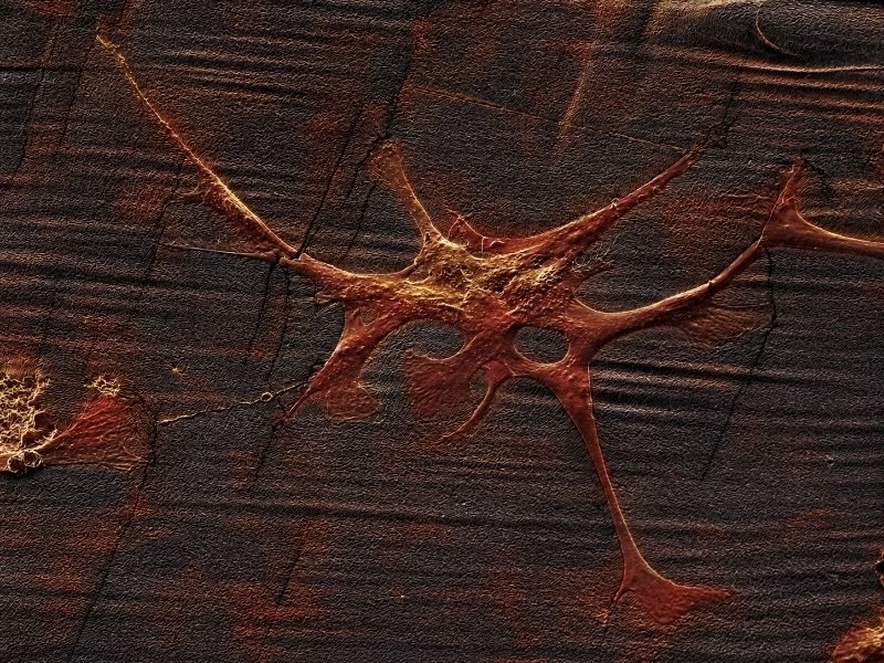
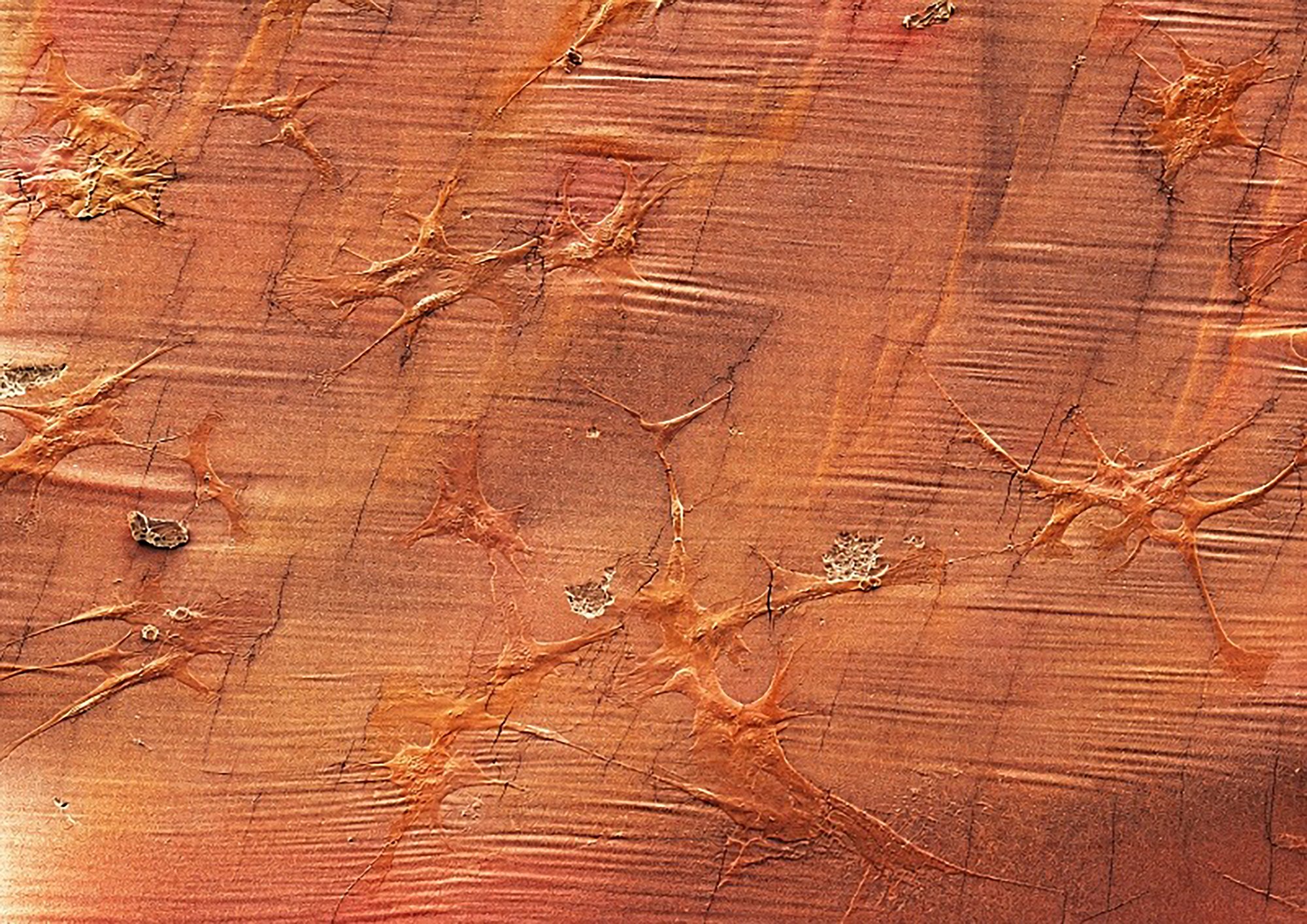
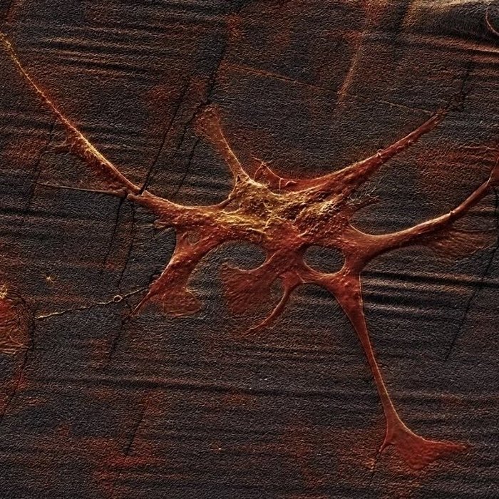
This work asks whether the geometry of a landscape can instruct the form that life takes — not only at the scale of memory and culture, but at the scale of the cell. Human neuronal-like cells were cultured on silicone substrates cast from a topographic model of the Himalayan mountain range, terrain drawn from personal memory of a place the artist has not stood on for years. Once differentiated, the cells were imaged using electron microscopy, producing photographs of neurites branching into forms that recall pine trees and mountain ridgelines — the same branching structure recurring across radically different scales, from the microscopic to the geological.
The work draws on the Chinese aesthetic philosopher Zhu Guangqian's account of three attitudes toward an old pine tree — utilitarian, scientific, and aesthetic — the third being the moment when viewer and object become one. This project proposes that the same convergence is visible under the microscope: the cell is not separate from the person who grew it, but expresses the same drive toward form, shaped by the same terrain.
The audience is invited to approach the work not as a literal illustration of a scientific hypothesis, but as visual evidence of something that actually happened — an experiment in which a physical, remembered landscape left its trace in living biological matter.
This project was developed with the support of the Francis Crick Institute, in collaboration with the Making Lab, the Electron Microscopy and the Cell Science Science Technology Platforms.
The substrate was fabricated by 3D-printing a resin negative mould of a Himalayan topographic model, then casting polydimethylsiloxane (PDMS) into the mould to produce a silicone scaffold bearing the mountain's terrain at approximately 18 × 36 mm scale. SH-SY5Y human neuroblastoma cells were seeded onto the PDMS scaffold, coated with Basement Membrane Extract to support neurite development. Cells were differentiated over a 10-day protocol using retinoic acid (10 μM) and BDNF (50 ng/mL).
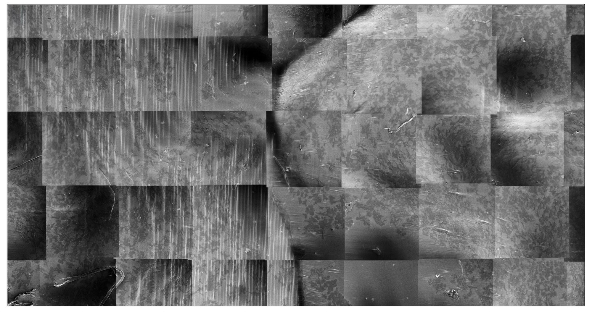
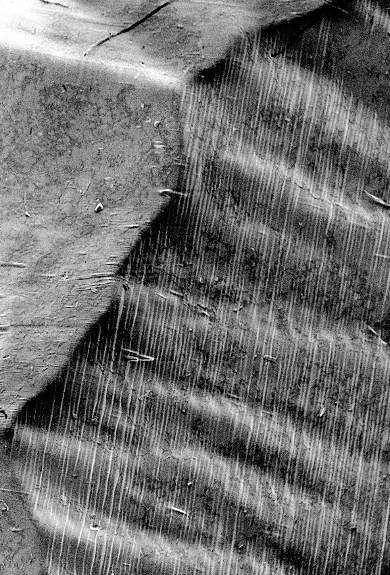
Once fixed, samples were imaged using two electron microscopy platforms at the Francis Crick Institute: a ThermoFisher Quanta/SECOM integrated light and scanning electron microscope, used for high-resolution close-up imaging of individual neurite morphology (including tilted-angle capture showing both cell and substrate topography in a single frame), and a Zeiss Gemini 460 SEM with Gatan 3View, used for serial block-face imaging across a 12-hour continuous session, producing a panoramic montage of over 200 individual image tiles stitched into a single view of the full scaffold surface.
The resulting dataset — pairing defined substrate geometry with observed neuronal morphology — represents a novel visual and biological record of environment-driven cell growth, distinct from standard flat-substrate cell culture imaging.
Xueqin Peng
Central Saint Martins, University of the Arts London, Francis Crick Institute, London
Xueqin Peng is an interdisciplinary practitioner working at the intersection of glassmaking, cell biology, and computational imaging. Prior to studying for an MA in Art and Science at Central Saint Martins, she spent over a decade in strategic operations within the technology industry. She completed a six-month research residency at the Francis Crick Institute, culturing neuronal-like cells on topographic silicone substrates and imaging them using electron microscopy. Her practice moves between material craft and biological experiment, asking how physical landscapes shape the forms life takes — from memory to matter.